
BioTechniques News
Beatrice Bowlby
Original story from the University of Oxford (UK).
Researchers have captured the step-by-step assembly of individual virus-like particles, revealing how simple molecular interactions can reliably build these complex biological structures.
Understanding how viruses assemble – and what can disrupt this process – could ultimately help researchers design novel antiviral treatments, as well as engineer vaccines and other therapies. Now, a study at the University of Oxford (UK) has provided a molecular-level view of how the protein shells associated with viruses can spontaneously and reliably assemble from their individual components, even when there are thousands of possible ways for these to fit together.
The study found that the process works rather like finding a route through a maze in which only the productive turns lock into place. Protein building blocks initially make weak, reversible connections, allowing unsuccessful arrangements to fall apart and be tried again. But when the proteins form particular closed structures, their multiple connections make them much more stable. These structures act as a small number of molecular waypoints, progressively funneling an otherwise highly complex process towards the completed particle.
A crucial moment occurs when five larger protein building blocks form a closed pentagonal ring, the first particularly stable structure in the pathway. After this step, fewer protein building blocks are needed to reach each new stable stage, allowing the assembly process to speed up.
Co-lead author, Roi Asor commented: “A virus has to solve an extraordinary construction problem. Its components somehow have to find the right arrangement among a huge number of possibilities, without a blueprint or machinery directing the process. We can now watch what happens molecule by molecule and see the physical rules that make it possible.”

Some of Europe’s most well-known historic manuscripts, including the York Gospels, have proven to be biological time capsules for uncovering the 3500-year history of the sheeppox virus (SPPV).
Capturing a molecular construction process
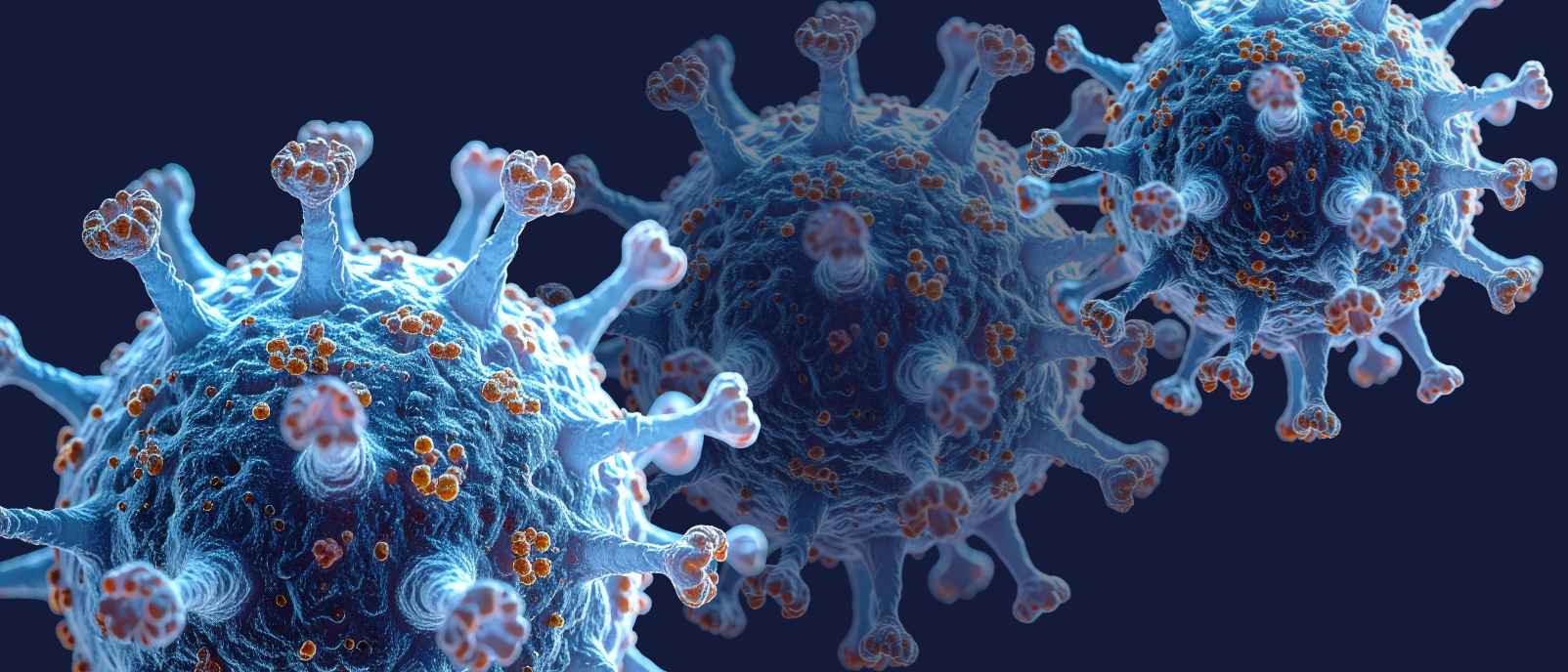
Viruses protect their genetic material inside precisely organized protein shells called capsids. These structures can contain tens or even thousands of protein components, yet in many viruses their building blocks spontaneously assemble into the correct shape.
Scientists have studied this process for decades, but observing it directly has proved extremely difficult. Crucial intermediate structures are scarce and often short-lived, meaning that conventional experiments can show researchers what is present before and after assembly without necessarily revealing the molecular steps connecting the two.
The Oxford team tackled this problem using an engineered virus-like particle made from 60 protein units. They combined mass photometry, a technology developed at Oxford that measures the mass of individual molecules by detecting the light they scatter, with a new method for confining individual molecules so that they could be observed continuously. This allowed the team to effectively ‘weigh’ a particle repeatedly as new protein components joined it, revealing its growth step by step.
Co-lead author Philipp Kukura shared: “Until now, much of our understanding of how these structures assemble has had to be reconstructed from snapshots or theoretical models. Being able to both quantify the underlying interactions and follow one particle as it grows changes that. We can see the important intermediate structures appear and deduce a detailed model of the construction process.”
The findings explain how an assembly process with an enormous number of theoretically possible routes can be channeled through a relatively small number of productive stages. They also reveal why the difficult initial step, known as nucleation, is followed by much faster growth once assembly is underway.
“The weak interactions give the system room to make mistakes. Most encounters don’t have to be successful: the components can separate and try again. But once enough of them come together in the right closed arrangement, the structure becomes stable and assembly can move forward. That combination of trial and error followed by locking in successful structures is what makes the process so reliable,” Asor explained.
Co-author Jack Tan noted: “The ability to understand these processes at the molecular level could have important applications in vaccine development and antivirals that disrupt viral assembly.”
More broadly, the principle could extend well beyond viruses. Self-assembly is fundamental to biology, underpinning structures ranging from the cellular skeleton to molecular compartments and protein complexes. The researchers say their approach provides a way to investigate these processes one molecule at a time.
This article has been republished from the following materials. Material may have been edited for length and house style. For further information, please contact the cited source. Our press release publishing policy can be accessed here.
The post Construction without a blueprint: the secret to how viruses assemble appeared first on BioTechniques.
Powered by WPeMatico