
BioTechniques News
Beatrice Bowlby
For the first time, the moment two DNA molecules zip together has been captured on camera, solving a mystery that has plagued scientists studying this fundamental biological process for more than 20 years.
For decades, the structural basis of DNA–DNA pairing has been an enigma; now, researchers from the University of Sheffield and the University of York (both UK) have combined atomic force microscopy and molecular dynamics simulations to provide the first direct visualization of the process in the presence of divalent ions. In doing so, they have finally revealed how DNA molecules overcome their electrical repulsion to lock together – a discovery that has implications f
or many key cellular processes, including chromosomal condensation and genome organization.
Recognition of homologous DNA sequences is essential in processes such as recombination. However, it presents a conundrum that has long puzzled scientists: why are DNA molecules able to pair up despite their identical negative charges? One leading theory to explain this contradiction is the ‘electrostatic DNA zipper’, or helical coherence, model. Originally proposed in 2001, the model posits that counterions bind to DNA grooves, creating alternating charge patterns that allow for electrostatic interlocking between the positively charged grooves of one duplex and the negatively charged backbone of another.
Although it’s been explored widely in the last 20 years, this hypothesis has never been proven, meaning the precise molecular mechanism of DNA pairing remains elusive and has yet to be visualized experimentally.
In a bid to change that, the team behind the latest study used high-resolution atomic force microscopy to image DNA–DNA pairing in a solution of divalent ions (below). They observed that major and minor grooves in paired double-stranded DNA matched up almost perfectly across the duplexes, suggesting that divalent ions align the grooves on adjacent DNA molecules as per the electrostatic zipper model.
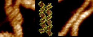
These results were supported by atomistic molecular dynamics simulations, which reproduced spontaneous self-assembly of DNA fragments when solvated by nickel cations. The DNA pairs interacted with each other in almost all simulations containing Ni2+, even if they were not homologous.
The simulations also demonstrated that some of the positively charged ions interact with both duplexes simultaneously at specific coordination sites within the minor groove, acting as a molecular bridge to stabilize the interaction.
Both the imaging and computational data revealed that coordination sites occur at non-random positions, indicating that the pairing mechanism varies depending on sequence and according to the specific divalent ion.
“To be able to directly visualize this long-hypothesized mechanism for the first time was incredible,” Thomas Catley (University of Sheffield), co-lead author of the study, remarked. “The advanced imaging techniques at our disposal have allowed us to uncover these key DNA interactions which have implications in many key cellular processes. It opens the door to studying other DNA interactions that, until now, have only existed as theory.”
“Hopefully, these programmable interactions could eventually help engineers design custom DNA structures for future biotechnology, such as DNA origami, and shed light on how DNA is actually packaged inside cells,” Catley added.
The post Zip it: scientists capture first-ever footage of two DNA strands locking together appeared first on BioTechniques.
Powered by WPeMatico