BioTechniques News
Tristan Free
Researchers have discovered a key transition in early embryonic development is facilitated by decreasing levels of a viral protein inserted into the DNA of our early animal ancestors.
Researchers at the Spanish National Cancer Research Center (CNIO; Madrid, Spain) have for the first time demonstrated that a protein encoded by genetic material from endogenous retroviruses (ERVs) plays a key role in early embryonic development. The team analyzed relationships between an ERV-encoded protein and key drivers of embryonic development in mice to establish this connection. Due to a great deal of conservation between embryonic development in different species of the animal kingdom, this research could provide relevant insight into the role of ERVs in human development.
Between 8 and 10% of our DNA originates from ERVs, deposited in early animal cells through retroviral infection and genome integration. These sections were long written off as ‘junk DNA’ but are now understood to have key regulatory roles. These regions have evolved symbiotically alongside our genomes and are known to contribute to the process of embryonic development in modern animals, a finely tuned sequence of events, engineered to ensure that development occurs correctly. How these steps are regulated and timed correctly remains mysterious to researchers.
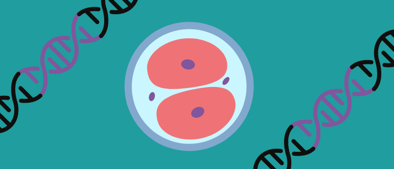
A key hallmark of embryonic development includes the progressive restriction of cell fates, as cells continually differentiate toward their final cell types. The first step of this restriction is the transition from totipotency to pluripotency. This reflects the loss of the ability of stem cells to differentiate into any cell type, including types that later make up extraembryonic tissues such as the placenta. Both cells in the two-cell stage of embryonic development are totipotent, but following division, the cells within a four-cell embryo are pluripotent.
In order to investigate this initial step and to better understand the regulatory mechanisms behind it, the research team deployed a suite of multiomic techniques – including immunofluorescence, chromatin immunoprecipitation sequencing analysis (ChIP-seq) and assay for transposase-accessible chromatin with sequencing (ATAC-Seq), alongside bioinformatics, genetic engineering co-immunoprecipitation assays and 3D cell culture methods – to study both in vitro and in vivo murine-based models of embryological development.
 Does junk DNA hold the key to aging?
Researchers have discovered a DNA region that drives the activity of the telomerase gene, a gene that influences the aging of certain cell types.
Using this approach, the researchers identified that a protein encoded by murine endogenous retrovirus-L (MERVL), MERVL-gag, is essential in this transition. MERVL-gag expression is high at the beginning of the two-cell stage but later decreases.
MERVL-gag regulates the timely loss of totipotency in mouse embryos through its interactions with the protein URI. Pluripotency-associated transcription factors are bound by URI, which prevents their proteasomal degradation. This increases the longevity of these factors and facilitates the transition to pluripotency. Deletion of URI is known to prevent embryonic development from occurring. MERVL-gag binding to URI maintains totipotency by allowing for the degradation of pluripotency factors in the early two-cell embryo. However, over time the levels of MERVL-gag decrease, allowing for pluripotency to arise.
The binding of URI to MERVL-gag in totipotent-like mouse embryonic stem cells, and to pluripotency factors in pluripotent mouse embryonic stem cells, was confirmed using co-immunoprecipitation. It is thought that the effects of the decreasing levels of MERVL-gag across the two-cell embryo stage allow for a gradual and well-timed change to pluripotency. “It’s a smooth transition. When there is a high expression of viral protein, there are fewer pluripotency factors; as ERV expression decreases, URI stabilizes such factors,” elaborates Sergio de la Rosa, the first author of this study.
Beyond enriching our understanding of the finely orchestrated steps of embryonic development, it is hoped that these findings will contribute to the field of regenerative medicine. For instance, this raises URI as a potential target in the development of stable totipotent stem cells, information that could also be applied to the development of programmable embryos.
The post Recycled “junk”: the viral protein essential in early embryonic development appeared first on BioTechniques.
Powered by WPeMatico